Recurrent Urinary Tract Infections in Men: Underlying Causes and Investigations
Learn why recurrent UTI in men requires thorough investigation, underlying causes, diagnostic approa

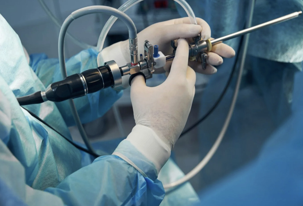
A cystoscopy uses a thin, lighted instrument called a cystoscope to provide a direct view of the urethra and the bladder’s interior lining. This commonly used diagnostic tool captures subtle details — such as minor colour changes, flat mucosal lesions, inflammation, or small growths — that imaging alone, such as ultrasounds or CT scans, may not reliably detect, particularly for early or superficial abnormalities. Depending on your specific clinical needs, your urologist will use either a flexible or rigid scope to aim for a thorough and accurate examination.
Urologists utilise cystoscopy to gain a clear, internal perspective when non-invasive tests like imaging or blood work provide an incomplete picture of urinary health. By allowing direct visualisation of the bladder and urethra, this procedure aims to help specialists identify potential underlying causes of persistent or complex symptoms that require more than just a surface-level scan.
Common reasons for this recommendation include:
Flexible cystoscopy utilises a thin, bendable scope and local anaesthetic gel to navigate the urethra with minimal discomfort, making it suitable for quick diagnostic checks in an outpatient setting. In contrast, rigid cystoscopy features a straight instrument with a wider working channel and improved irrigant flow, making it better suited for surgical interventions such as tumour removal or stone treatment.
While rigid scopes traditionally offered high image quality, both flexible and rigid types have demonstrated equivalent accuracy in identifying bladder tumours. Your urologist will choose the instrument based on your specific needs, typically opting for the flexible approach for routine surveillance and the rigid version for more complex therapeutic procedures.
Proper preparation is designed to support patient safety and aims to minimise the risk of complications such as bleeding or infection.
If you take blood-thinning medications, your doctor will provide a specific plan to adjust or temporarily stop them based on your individual health risks. Most diagnostic patients can eat normally, but those undergoing sedation or general anaesthesia must follow strict fasting instructions for several hours. Always consult your healthcare provider before making any changes to your prescribed medication routine.
A urine sample is typically collected beforehand to check that no active infection is present, as performing the procedure during a UTI could spread bacteria into the bloodstream. If an infection is detected, the cystoscopy is usually postponed until a course of antibiotics has cleared the infection. Decisions about prophylactic antibiotics are made on an individual basis by your urologist, taking into account your overall health, immune status, and procedure type. Routine antibiotic prophylaxis is not universally recommended and is guided by current clinical protocols — always follow the specific advice given by your healthcare team.
The procedure follows a standardised clinical process designed to help support patient comfort while providing the urologist with a clear view of the urinary tract.
For a flexible examination, you will lie on your back while a local anaesthetic gel is applied to the urethra to numb and lubricate the area. If a rigid scope is used, you may receive spinal or general anaesthesia and will be placed in the lithotomy position with your legs elevated. These measures are designed to help you remain comfortable and still, allowing the medical team to safely navigate the instrument.
As the urologist gently advances the scope, you may feel some pressure or the sensation of needing to urinate. A sterile irrigating fluid — typically sterile saline or sterile water, selected based on the type of procedure being performed — is then introduced into the bladder through the cystoscope to gently expand its walls, creating the necessary space for a detailed visual inspection. Practising slow, deep breaths during this stage helps relax your pelvic muscles and manage the temporary urge to void.
The urologist methodically rotates the scope to inspect every surface of the bladder, including the dome, side walls, and the trigone — the triangular region on the bladder floor where the ureteral openings are located. They carefully observe the ureteral openings for urine flow and pay close attention to the bladder neck and urethra for any signs of pathology. Throughout the process, the doctor documents the health of the lining and may take photographs or biopsies of any suspicious areas.
If suspicious areas appear, your urologist may collect tissue samples using small forceps passed through the cystoscope’s working channel. During this tissue collection, you may feel brief pinches, and if the biopsy site is cauterised to control bleeding, a brief burning sensation may also be experienced. The bladder lining has limited pain sensation, but individual experiences vary. Collected tissue goes to a pathology laboratory for microscopic examination. Results typically take several days.
Minor bleeding from tissue collection sites is normal and self-limiting. More significant procedures, such as tumour removal, are performed through the rigid cystoscope using specialised instruments.
Treatment of an enlarged prostate — such as transurethral resection of the prostate (TURP) — is performed using a related but distinct instrument called a resectoscope, which incorporates a cutting wire loop and electrosurgical capability. These use specialised instruments, including electrocautery (devices that use heat to cut or seal tissue), lasers, or mechanical resection devices.
The examination concludes with bladder drainage and gentle scope withdrawal. You can usually stand and walk immediately after flexible cystoscopy.
It is common to experience a mild burning sensation or pink-tinged urine during your first few voids as the anaesthetic gel wears off and minor irritation subsides. Increasing your fluid intake helps flush the urinary system and dilutes the urine to reduce this localised discomfort. Most of these minor side effects should naturally clear up within a day or two of the examination.
Patients undergoing a diagnostic flexible cystoscopy under local anaesthetic can typically resume normal activities relatively quickly, but should confirm with their doctor before driving, as individual recovery varies. They should avoid strenuous exercise for 24 hours. If your procedure involved sedation or general anaesthesia, you must arrange for a responsible adult to drive you home. For these cases, you should also avoid operating machinery or making major decisions for at least a full day.
Your urologist will often provide an immediate preliminary report, but definitive answers regarding tissue samples will require several days of laboratory analysis.
Ask your urologist specific questions about what they expect to find. Ask what they’ll do if they discover abnormalities. Knowing that many cystoscopies reveal either normal findings or manageable conditions provides realistic reassurance.
Arrange your schedule to allow rest after the procedure if needed. While many patients resume normal activities quickly, having flexibility reduces stress. Bring reading material or entertainment for any waiting periods.
Focus on slow, regular breathing throughout the procedure. Tension in pelvic floor muscles increases discomfort during scope insertion. Some patients find it helpful to imagine breathing “through” the sensation rather than fighting it.
Communication with your medical team matters. Inform them immediately if you experience sharp pain. This differs from the normal pressure sensation and may indicate a need for adjustment. Discomfort during certain portions of the examination is usually brief.
How painful is cystoscopy without sedation?
Flexible cystoscopy with local anaesthetic gel causes discomfort rather than true pain for many patients. The sensation resembles needing to urinate urgently, combined with pressure. Brief stinging occurs during scope passage through the sphincter. The examination portion feels like bladder fullness. Individual experience varies — while many patients find the procedure uncomfortable but tolerable, some may experience more significant discomfort, and it is entirely appropriate to communicate this to your care team.
Can cystoscopy detect bladder cancer?
Cystoscopy can directly visualise the bladder lining and identify small tumours. It is a commonly performed method for bladder cancer detection and surveillance in patients who may be at risk. However, some flat cancers (carcinoma in situ, where abnormal cells are present on the bladder surface) may be difficult to see with standard white light cystoscopy. These require special techniques like blue-light cystoscopy for more reliable detection.
How long does a cystoscopy take?
The cystoscopy examination itself typically takes 5 to 10 minutes for a straightforward diagnostic flexible procedure. Including preparation, the full appointment usually takes 15 to 30 minutes. Rigid cystoscopy with tissue sampling or therapeutic interventions takes longer, depending on complexity. Recovery room time adds to the total visit duration if sedation or general anaesthesia is used.
When can I return to work after a cystoscopy?
Many patients return to desk work the same day or the following morning after flexible cystoscopy. Physical labour may require a day or two off due to minor urinary discomfort. Rigid cystoscopy under general anaesthesia typically requires a short recovery period before returning to work.
Do I need someone to drive me home?
If you had a flexible cystoscopy under local anaesthetic only, many patients are able to drive themselves home — but you should confirm this with your doctor beforehand, as individual recovery varies. Any procedure involving sedation or general anaesthesia requires a responsible adult to accompany you home, and you should not drive for at least 24 hours after sedation.

Former Director of Endourology (Urinary stone service) Singapore General Hospital 2016 to 2023
With more than 20 years experience as a certified Urologist, Dr Nor Azhari specializes in treating a wide range of kidney, bladder and prostate conditions as well as disorders of the male reproductive organs. He offers minimally invasive treatment options and provides same-day appointments for convenience.
For urgent or same day appointment requests, please call our hotline.
Learn why recurrent UTI in men requires thorough investigation, underlying causes, diagnostic approa
 Optimized by Seraphinite Accelerator