Fraud Blocker

Same Day Appointments Available

Understanding Kidney Obstruction Treatment in Singapore

If you have been told that your kidney is “blocked” or that urine is not draining properly, you are dealing with what urologists call a ureteric stricture or pelviureteric junction (PUJ) obstruction. These conditions affect the kidney and ureter, the tube that carries urine from the kidney to the bladder. In Singapore, urological intervention focuses on relieving that obstruction and protecting your kidney’s long-term function.

doctor img
Dr. Nor Azhari Bin Mohd Zam

MBBS (NUS)|MRCS (Edin.)|MMed Surgery (NUS)|FAMS (Urology)

Illustration comparing a normal urinary system with a ureteropelvic junction obstruction. Understanding Kidney Obstruction Treatment in Singapore

What is Kidney Obstruction?

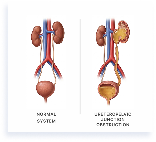
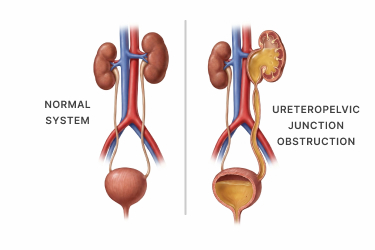
A kidney obstruction is more than a structural problem on a scan. You may have been living with persistent flank discomfort, repeated urinary tract infections, or the knowledge that urine is pooling where it should not be. Many patients initially dismiss these signs as “just a backache” or blame recurring infections on something else entirely.

A specialist consultation bridges those everyday complaints with the urologist’s core objective: restoring normal urinary drainage to protect your kidney from progressive, and often irreversible, functional decline.

How Is Kidney Obstruction Identified?

Many patients notice something is wrong long before they receive a formal diagnosis. You might describe it as a dull ache in your back or side that simply will not go away. Perhaps an ultrasound ordered for another reason revealed swelling in your kidney. Or maybe you have been battling urinary tract infections that keep returning despite completing full courses of antibiotics.

These everyday observations matter. They are often the very clues that prompt your urologist to investigate further. Translating these symptoms into a confirmed diagnosis of ureteric stricture or PUJ obstruction requires a structured clinical assessment.

Physical Examination and Initial Assessment

Your urologist will start with a thorough history and physical examination. They will ask about the nature of your pain, its location, how long it has persisted, and whether you have experienced fevers, nausea, or changes in your urinary habits.

Tenderness over the flank area during examination can raise suspicion, but physical findings alone are rarely enough to confirm an obstruction. That is why imaging plays such a critical role.

Imaging Studies

Ultrasonography is typically the first-line investigation. It is non-invasive, widely available, and effective at detecting hydronephrosis, the swelling of the kidney caused by urine backup. If the ultrasound suggests an obstruction, your specialist will usually recommend more detailed imaging.

CT urography provides a comprehensive, cross-sectional view of the kidneys, ureters, and bladder. It can pinpoint the exact location and potential cause of the blockage, whether it is a narrowing within the ureter itself or compression from an external structure. CT urography is also useful for ruling out conditions that mimic obstruction, such as kidney stones or infections.

Functional Assessment

Identifying where the blockage is located is only part of the picture. Your urologist also needs to know how well each kidney is actually working. This is where nuclear renal scintigraphy comes in.

A MAG3 or DTPA renogram involves injecting a small amount of radioactive tracer into your bloodstream and tracking how efficiently each kidney filters and drains it. This study provides two essential pieces of information:

  • Split renal function: how much work each kidney is contributing
  • Drainage pattern: whether urine is flowing freely or being held up

Together, these imaging and functional tests form the diagnostic foundation that guides your treatment pathway.

What Causes Kidney Obstruction to Develop?

Understanding why your kidney became obstructed helps you make sense of your diagnosis. The causes generally fall into two broad categories: conditions present from birth and conditions that developed over time.

Congenital Causes

Some patients have lived with a predisposition to kidney obstruction since birth, even if symptoms only appeared in adulthood. The most common congenital cause is an intrinsic PUJ obstruction. This occurs when the segment of the ureter where it connects to the kidney does not contract normally.

Instead of propelling urine downward in rhythmic waves, this aperistaltic segment (a portion of the ureter that has lost its peristaltic contractions) acts as a bottleneck, preventing efficient drainage.

Another congenital cause involves aberrant crossing vessels, blood vessels that cross over the ureter near the PUJ and physically compress it. In some people, this compression only becomes clinically significant later in life, particularly during periods of high fluid intake.

Acquired Causes

Acquired ureteric strictures develop as a result of injury, inflammation, or changes to surrounding tissues. Common acquired causes include:

  • Scarring from prior surgical instrumentation: Procedures involving the ureter, such as ureteroscopy for kidney stone removal, can occasionally lead to scar tissue that narrows the ureteric lumen over time.
  • Radiation therapy directed at the pelvis, often for gynaecological or colorectal cancers, can cause delayed inflammation and fibrosis of the ureter.
  • External compression from masses, enlarged lymph nodes, or retroperitoneal fibrosis [scarring behind the abdominal lining] can also obstruct urine flow from outside the ureter wall.
How Obstruction Affects the Kidney

Regardless of the cause, the downstream effect is similar. When urine cannot drain efficiently, pressure builds within the kidney’s collecting system, leading to progressive hydronephrosis. Over time, this sustained pressure damages the renal parenchyma [the functional tissue responsible for filtering your blood].

Left unaddressed, this damage can become permanent, leading to irreversible loss of kidney function on the affected side. The kidney often does not send clear distress signals until significant damage has already occurred, which is why early identification matters.

Different Types and Stages of Kidney Obstruction

Not all kidney obstructions are the same. The type, location, and severity of the blockage directly influence how your specialist plans your care.

Classification by Location

The two primary types are distinguished by where the obstruction sits:

  • PUJ obstruction occurs at the junction where the renal pelvis meets the ureter. This is the most common site for congenital obstruction.
  • Ureteric stricture can occur anywhere along the length of the ureter and is more commonly associated with acquired causes such as prior surgery, radiation, or inflammation.

Intrinsic Versus Extrinsic Obstruction

Your specialist will also determine whether the blockage originates within the wall of the ureter itself (intrinsic) or from pressure applied by a structure outside the ureter (extrinsic). This distinction affects which treatment approaches are most suitable.

Partial Versus Complete Obstruction

The degree of blockage matters significantly:

  • A partial obstruction may allow some urine to pass. You might notice occasional flank pain after drinking a lot of water, which settles on its own.
  • A complete obstruction blocks urine flow entirely, often presenting as severe pain accompanied by fever. When combined with infection, this is considered a urological emergency.

Unilateral Versus Bilateral

Most kidney obstructions affect one side only (unilateral). Bilateral obstruction, in which both kidneys are affected, poses a more urgent threat because it compromises your body’s overall ability to produce and drain urine.

Grading Severity

Clinical grading relies on two key measurements:

  • The degree of hydronephrosis on imaging, which shows how swollen the kidney has become
  • The differential renal function on renography, which reveals how much filtering capacity the affected kidney has retained

Together, these parameters help the urologist assess urgency and determine whether surveillance, minimally invasive intervention, or reconstructive surgery is the most appropriate course of action.

When to Seek Specialist Advice

If you have been managing symptoms with pain relievers, increasing your water intake, or relying on your general practitioner to treat recurring infections, it is important to recognise when those strategies have reached their limit.

Seek a specialist urological evaluation if you experience any of the following:

  • Persistent or worsening hydronephrosis on follow-up imaging
  • Declining split renal function demonstrated on renography
  • Recurrent urinary tract infections despite appropriate antibiotic treatment
  • Unrelenting flank pain that disrupts your sleep, work, or daily activities

Drinking more fluids will not resolve a structural obstruction. Increasing fluid intake when a blockage is present may actually worsen pressure within the kidney. Over-the-counter painkillers may mask symptoms without addressing the underlying cause.

One scenario demands immediate specialist attention: when infection coexists with obstruction. An infected, obstructed kidney, sometimes called pyonephrosis, can lead to sepsis if drainage is not established promptly. If you develop fever, chills, and flank pain together, seek urgent medical care without delay.

Timely referral to a urologist helps ensure that a treatable condition does not silently progress to the point where kidney function is permanently lost.

Experiencing recurring infections or flank pain?

A structured urological assessment can determine whether a structural obstruction is present and establish a clear diagnostic pathway before kidney function is compromised.

Your Treatment Path

The appropriate approach to treating kidney obstruction depends on several factors: the severity of the blockage, your kidney’s function, your anatomy, and the impact of symptoms on your daily life. Your urologist will tailor a management plan based on these clinical variables, with the overarching goal of relieving the obstruction and preserving as much renal function as possible.

Mild Kidney Obstruction

When imaging reveals low-grade hydronephrosis and kidney function remains well preserved, your specialist may recommend active surveillance rather than immediate procedural intervention. This does not mean your condition is being ignored. It means the clinical picture does not yet justify the risks of surgery.

Active surveillance involves serial imaging and renography at regular intervals, typically every six to twelve months, to track whether hydronephrosis is stable or progressing and whether your kidney’s filtering capacity is holding steady.

Triggers that would prompt escalation to intervention include:

  • A measurable drop in split renal function
  • Worsening hydronephrosis
  • New onset of symptoms
  • Development of urinary tract infections
Moderate Kidney Obstruction

When there is clear evidence of impaired renal drainage, progressive hydronephrosis, or a significant symptom burden, active intervention becomes clinically appropriate. Several minimally invasive options may be considered:

  • Endopyelotomy: an incision through the narrowed segment to widen the passage
  • Balloon dilatation: a small inflatable device used to stretch the stricture open
  • Ureteric stenting: a thin tube placed within the ureter to hold it open and allow urine to drain while the area heals or while planning definitive treatment

The choice of technique depends on the characteristics of the stricture, its length, its location, and whether it is intrinsic or extrinsic. Patient factors, such as body habitus, prior surgical history, and overall health, also play a role. Dr Azhari works with patients to select the approach that aligns with their clinical needs, discussing the potential risks and limitations of each option beforehand.

Severe Kidney Obstruction

In cases of high-grade or complete obstruction, significant renal functional compromise, infected hydronephrosis, or failure of previous endoscopic management, reconstructive surgical approaches become necessary.

For PUJ obstruction, pyeloplasty, the surgical removal of the obstructed segment and reconnection of the renal pelvis to the ureter, is considered the standard reconstructive procedure. This can be performed through open, laparoscopic, or robotic-assisted approaches.

For extensive ureteric stricture disease, techniques such as ureteric reimplantation or substitution procedures using other body tissues may be required. Where a kidney has lost all meaningful function, as confirmed by thorough specialist evaluation, nephrectomy (surgical removal of the kidney) may be considered to prevent ongoing complications such as chronic infection or pain.

All surgical decisions carry inherent risks, which must be weighed carefully against the potential clinical benefit in a detailed, transparent discussion with your urologist.

Has a treatment plan been recommended for your kidney obstruction?

Understanding the available procedural and surgical pathways allows patients to make informed decisions aligned with their clinical findings and recovery goals.

Clinical Goals and Safety

The overarching clinical goals of treating kidney obstruction are to preserve functioning renal tissue, restore the natural flow of urine, and prevent irreversible nephron loss. When intervention occurs at an appropriate time, many patients may experience improvement in both symptoms and measured kidney function.

What Influences Outcomes

Results vary from person to person. Several factors influence how your kidneys respond to treatment:

  • Duration of obstruction before intervention: A kidney obstructed for months or years may have sustained damage that cannot be fully reversed, even after drainage is restored.
  • Baseline renal function: If your kidney was already underperforming at diagnosis, the ceiling for recovery may be lower.
  • Patient comorbidities: Conditions such as diabetes, hypertension, or chronic kidney disease can affect healing and long-term renal health.
  • The specific cause of the stricture: Congenital obstructions managed early may carry a more favourable outlook compared with strictures caused by radiation or extensive fibrosis.

Safety Considerations

Both endoscopic and surgical interventions are performed within established safety frameworks. Minimally invasive techniques generally offer smaller incisions, reduced blood loss, and shorter hospitalisation. Open reconstructive procedures remain appropriate for complex cases where minimally invasive access is not feasible or has previously been unsuccessful.

Every procedure carries potential complications, including bleeding, infection, stricture recurrence, or the need for further intervention. Dr Azhari reviews these risks in detail during pre-operative counselling so that you can make an informed decision.

Can Kidney Obstruction Be Prevented?

While not all kidney obstructions can be prevented, there are meaningful steps that may reduce the risk of developing or worsening the condition.

  • Early Screening for At-Risk Patients

    If you were born with a known urinary tract anomaly, have undergone previous urological surgery, or have received pelvic radiation therapy, regular specialist screening can detect early signs of obstruction before symptoms appear. Ultrasound surveillance and periodic renal function assessments allow your urologist to identify subclinical hydronephrosis and intervene before permanent damage occurs.

  • Prompt Management of Related Conditions

    Kidney stones left untreated or requiring repeated instrumentation can contribute to ureteric scarring. Prompt and appropriate management of urinary calculi may reduce this risk. Careful surgical technique during any procedure involving the ureter is equally important, as minimising trauma to the ureteric wall helps prevent stricture formation.

  • Long-Term Urological Surveillance

    Patients with a history of PUJ obstruction, ureteric surgery, or pelvic radiation benefit from long-term urological follow-up to catch any recurrence early. Surveillance is a proactive clinical strategy designed to protect your kidney over the years ahead, not a sign that something has gone wrong.

Dr Azhari advises patients with predisposing conditions to maintain a consistent surveillance schedule, as even subtle changes on imaging or function testing can signal the need for timely intervention.

Frequently Asked Questions

What does a blocked kidney feel like?

Many patients describe a dull, persistent ache in the back or side, often below the ribs. Some experience pain that worsens after drinking large amounts of fluid. Others have no pain at all and only discover the obstruction through incidental imaging.

Can a blocked kidney resolve without treatment?

In most cases, a true structural obstruction, such as a ureteric stricture or PUJ obstruction, will not resolve on its own. Temporary conditions like swelling after passing a kidney stone may improve naturally, but confirmed strictures typically require specialist management.

Is kidney obstruction the same as kidney stones?

No. Kidney stones are solid mineral deposits that can cause a temporary blockage as they pass through the ureter. Kidney obstruction from a stricture or PUJ narrowing is a structural problem that persists regardless of whether stones are present. Stones and strictures can, however, coexist.

Will I need surgery, or can it be treated with medication?

There is no medication that can open a ureteral structural blockage. Treatment depends on severity. Mild cases may be monitored, while moderate to severe cases typically require procedural or surgical intervention. Your specialist will recommend the most appropriate course of action based on your individual findings.

How do doctors check if my kidney is still working?

A nuclear renal scan, known as a MAG3 or DTPA renogram, measures how well each kidney filters and drains. Blood tests for creatinine and estimated glomerular filtration rate also provide information about overall kidney health.

Can the obstruction come back after treatment?

Recurrence is possible, particularly with certain stricture types or after endoscopic procedures. This is one of the key reasons why long-term follow-up imaging and function testing are considered a clinical standard after treatment.

Is this condition dangerous if left untreated?

Yes. Untreated obstruction can lead to progressive kidney damage, chronic infections, and in severe cases, permanent loss of the affected kidney’s function. An infected, obstructed kidney can become a medical emergency.

Can children have kidney obstruction?

Yes. PUJ obstruction is one of the most common congenital urological conditions detected in children, often identified on prenatal ultrasound. Early specialist evaluation helps determine whether the child needs intervention or can be safely monitored.

How long does treatment typically take?

This varies widely. A stent placement may take under an hour, while a reconstructive pyeloplasty may take two to three hours. Your specialist will discuss the expected procedure duration and hospital stay during your consultation.

Long-Term Renal Health and Clinical Success

Clinical success after kidney obstruction treatment is not defined by a single procedure. It is measured by the sustained stabilisation of your kidney and ureter over months and years, confirmed through serial imaging and renal function monitoring.

Long-term follow-up is a clinical standard, not an optional recommendation. With structured oversight from an experienced urologist, patients can transition from active treatment to a proactive surveillance plan designed to protect their kidney function over time.

To discuss your kidney health or arrange a specialist review, consider booking a consultation to understand your diagnostic findings and the options available to you.

Concerned about kidney obstruction or long-term renal health in Singapore?

Dr Azhari provides diagnostic clarity and structured follow-up to help protect kidney function and prevent irreversible decline.

Dr. Nor Azhari Bin Mohd Zam

Dr. Nor Azhari Bin Mohd Zam

MBBS (NUS)|MRCS (Edin.)|MMed Surgery (NUS)|FAMS (Urology)

With more than 15 years experience as a certified Urologist, Dr Nor Azhari specializes in treating a wide range of kidney, bladder and prostate conditions as well as disorders of the male reproductive organs.

He offers minimally invasive treatment options and provides same-day appointments for convenience.

  • Skilled urologist and kidney stone surgeon.
  • Director of Endourology (Urinary stone service) at Singapore General Hospital – 2016 to 2023.
  • Recognized with the College of Surgeons Gold Medal and Singapore Urological Association Book prize.
  • Minimally invasive treatment options available (non-invasive and endoscopic/key-hole techniques).

Make an Enquiry

For urgent or same day appointment requests, please call our hotline.

    Full Name*

    Email Address*

    Phone Number*

    Your Message*

    For Faster Response, call us!

    +65‎ 6334‎ 1486

    Our Clinic Locations

    Asian Healthcare Specialists (Novena)

    Mount Elizabeth Novena Specialist Centre
    38 Irrawaddy Road
    #08-41 Singapore 329563

    Asian Healthcare Specialists (Alvernia)

    Mount Alvernia Hospital
    820 Thomson Road Medical Centre Block A #05-03 Singapore 574623