Curvature of the Penis: Understanding Peyronie’s Disease and Treatment Options
Learn about Peyronie's disease treatment options in Singapore, from oral medications to surgery. Und


Why does blood appear in urine without pain or infection? This bleeding occurs when tumours growing on the bladder lining erode blood vessels, releasing red blood cells into urine. The bladder’s inner surface, lined with transitional epithelial cells called urothelium (the protective layer of cells that line the bladder), can develop abnormal growths. These range from small, benign papillomas to aggressive carcinomas (malignant tumours) that penetrate deeper tissue layers.
Bladder tumours develop through genetic mutations (changes in the DNA of urothelial cells). These are often triggered by prolonged exposure to carcinogens (cancer-causing substances) filtered through the kidneys. These mutations accumulate over the years. The bladder’s role as a storage organ means that harmful substances in urine remain in contact with the bladder wall for extended periods. This increases the likelihood of mutation compared to organs where urine passes through quickly.
Haematuria—blood in urine—presents in two distinct forms that require different detection methods. Gross haematuria produces visibly pink, red, or cola-coloured urine that patients notice immediately. Microscopic haematuria (blood in urine that’s invisible to the naked eye) remains invisible. It’s detectable only through urinalysis (a laboratory test of urine) that shows elevated red blood cell counts. Both warrant investigation, though visible blood typically prompts faster medical consultation.
The bleeding pattern in bladder tumours is intermittent. Blood may appear for several days, disappear completely for weeks, then return. This intermittency leads some people to dismiss the symptom when bleeding stops, assuming the problem has resolved itself. Tumour surfaces bleed when disturbed by bladder contractions during urination or when vessels erode. Temporary clotting can pause bleeding until the next disruption occurs.
Blood clots may accompany haematuria. These sometimes cause discomfort during passage or temporary urinary obstruction. The colour intensity doesn’t reliably indicate tumour severity. Small tumours with rich blood supply can produce dramatic bleeding while larger tumours may cause only subtle discolouration. Any blood in urine, regardless of amount or duration, warrants urological evaluation to determine its source. Your doctor can use diagnostic tools to identify the cause and determine appropriate next steps.
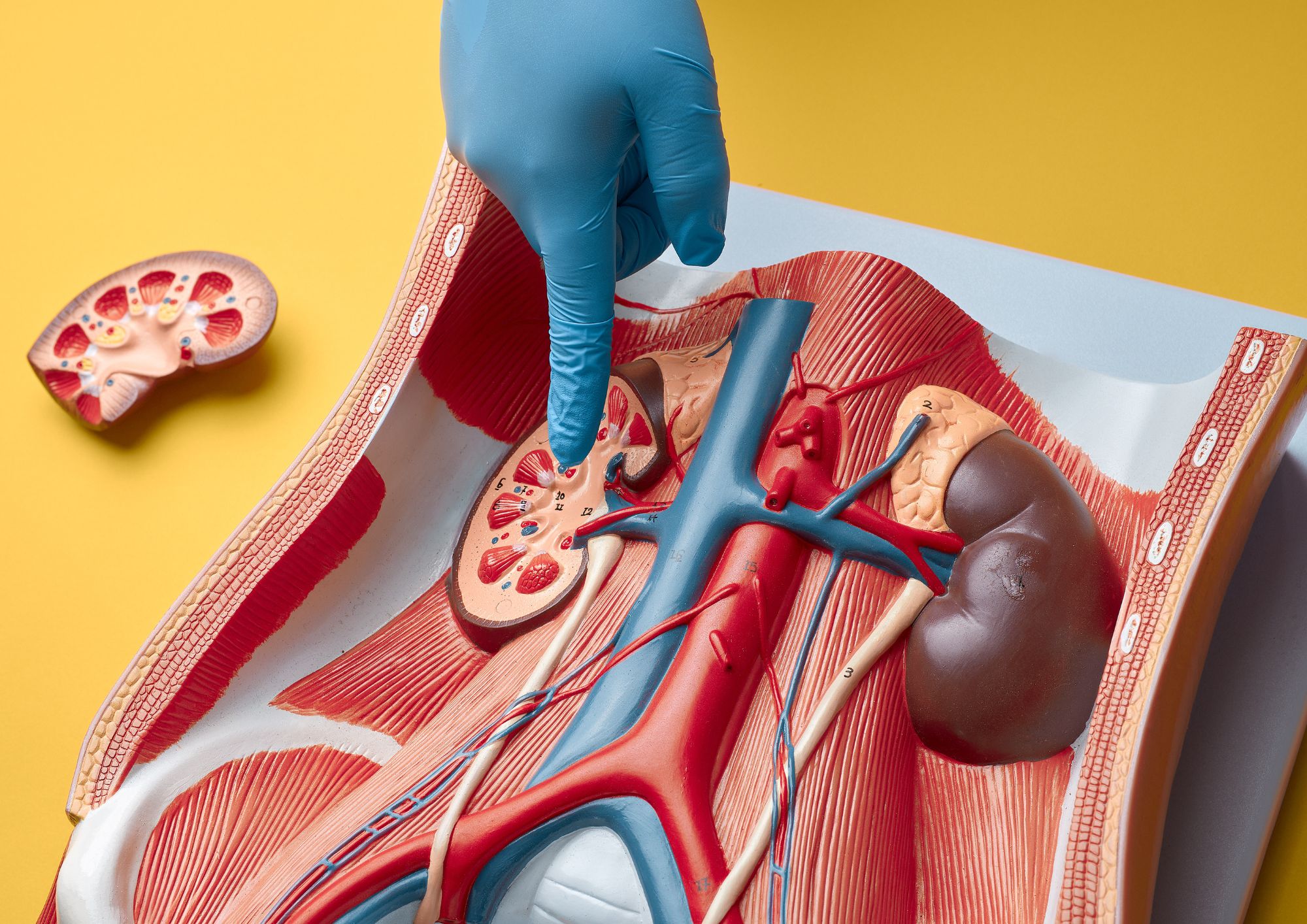
Bladder tumours alter standard voiding patterns through several mechanisms. Tumours occupying the bladder space reduce functional capacity. This creates urgency and frequency as the bladder reaches fullness sooner. Growths near the bladder neck (the opening where urine leaves the bladder) or ureteral openings (where the tubes from the kidneys connect to the bladder) can partially obstruct urine flow. This causes hesitancy or a weak stream.
Irritative symptoms—urgency (the sudden, strong need to urinate), frequency (needing to urinate more often than usual), and discomfort during urination—sometimes precede visible bleeding. Carcinoma in situ (a flat, high-grade tumour spreading across the bladder surface without forming a distinct mass) can cause intense irritation despite its non-bulky nature. Patients describe sensations similar to urinary tract infections, but without bacterial growth on culture.
Nocturia (waking multiple times nightly to urinate) may indicate reduced bladder capacity from tumour growth. While nocturia has many causes, including prostate enlargement in men, new-onset nocturia combined with other urinary changes warrants investigation. The combination of irritative and obstructive symptoms occurring together, especially in someone without previous urinary complaints, raises suspicion for bladder pathology.
Early bladder tumours typically cause no pain. Pain development usually indicates either infection, obstruction, or advanced disease involving deeper structures. Discomfort during urination without infection may signal tumour irritation of the bladder trigone—the sensitive triangular area between the ureteral openings and the urethra.
Flank pain (discomfort in the side of the body between the ribs and hip) can occur when tumours obstruct the ureteral openings. This causes urine backup and kidney swelling. This obstruction develops gradually as tumours grow. It sometimes reaches a significant size before causing noticeable discomfort. Unilateral flank pain (pain on one side only) with haematuria particularly suggests ureteral involvement requiring prompt imaging.
Pelvic pain or pressure sensations may indicate locally advanced disease extending beyond the bladder wall. Tumours invading surrounding tissues contact nerve endings absent from the bladder’s inner lining. This produces new pain patterns. Bone pain, weight loss, and fatigue suggest metastatic spread (cancer that has spread to other parts of the body) and warrant urgent comprehensive evaluation.
Smoking represents a significant modifiable risk factor for bladder cancer—carcinogens from tobacco concentrate in urine, bathing bladder cells in harmful chemicals for hours daily. Former smokers retain an elevated risk for years after quitting, though the risk decreases progressively with time since cessation. Current and former smokers should maintain heightened awareness of urinary changes.
Occupational exposures affect workers in specific industries. Aromatic amines (a group of industrial chemicals) used in dye manufacturing, rubber production, leather processing, and textile industries directly damage urothelial DNA. Workers in painting, hairdressing, and printing also face elevated exposure risks. Symptoms appearing years or decades after exposure reflect the long latency between carcinogen exposure and tumour development.
Previous pelvic radiation for other cancers increases bladder cancer risk in the treatment field. Cyclophosphamide chemotherapy (a cancer treatment drug), used for various malignancies and autoimmune conditions, produces a metabolite (a chemical produced when the body breaks down the drug) that is toxic to the bladder lining. Chronic bladder irritation from repeated infections, long-term catheter use, or bladder stones creates ongoing cellular turnover, increasing mutation opportunities.
? Did You Know?
The bladder lining completely replaces itself regularly through continuous cell division. This high turnover rate allows healing from minor injuries. However, it also provides more opportunities for DNA copying errors that initiate cancer development.
Urological assessment begins with a detailed history. This focuses on bleeding characteristics, urinary pattern changes, pain, and exposure to risk factors. Physical examination includes abdominal palpation (pressing on the abdomen to feel for abnormalities) for bladder distension. In men, it includes digital rectal examination (the doctor inserts a gloved finger into the rectum to feel the prostate) to assess prostate involvement.
Urine cytology (a laboratory test that examines cells in urine under a microscope) examines voided urine for abnormal cells shed from tumours. High-grade tumours shed distinctive cells that cytopathologists (specialists who study cells to diagnose disease) identify readily. Low-grade tumours may shed normal-appearing cells despite their presence. Urine cytology offers high specificity—positive results strongly suggest malignancy. However, it shows variable sensitivity depending on tumour grade.
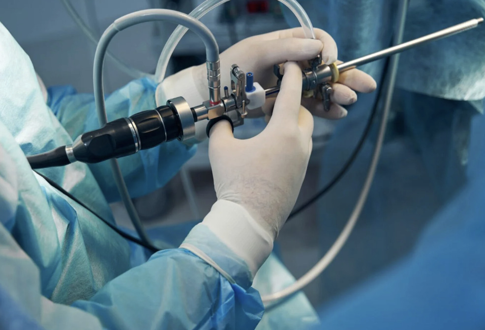
Cystoscopy (a procedure in which a thin camera is inserted through the urethra to visualise the bladder) provides direct visualisation of the bladder. This procedure allows urologists to inspect the entire bladder surface. They identify tumours, suspicious areas, and other abnormalities. Flexible cystoscopy performed under local anaesthesia (numbing medication applied to the area) in clinic settings offers initial screening. Rigid cystoscopy under anaesthesia permits biopsy (removal of a small tissue sample for testing) and treatment.
CT urography (a specialised imaging scan using X-rays and contrast dye to create detailed pictures of the urinary tract) images the entire urinary tract. It can detect bladder tumours while also evaluating kidneys and ureters for synchronous disease (cancer appearing in multiple locations at the same time). Contrast dye highlights the urinary system, revealing filling defects caused by tumours. This imaging method has largely replaced intravenous urography for evaluation of haematuria due to its ability to provide greater detail and additional information about surrounding structures.
⚠️ Important Note
A single normal cystoscopy doesn’t exclude bladder cancer if symptoms persist. Carcinoma in situ may appear as subtle redness rather than obvious tumour. Small growths can hide in bladder folds. Persistent haematuria or irritative symptoms warrant repeat evaluation.
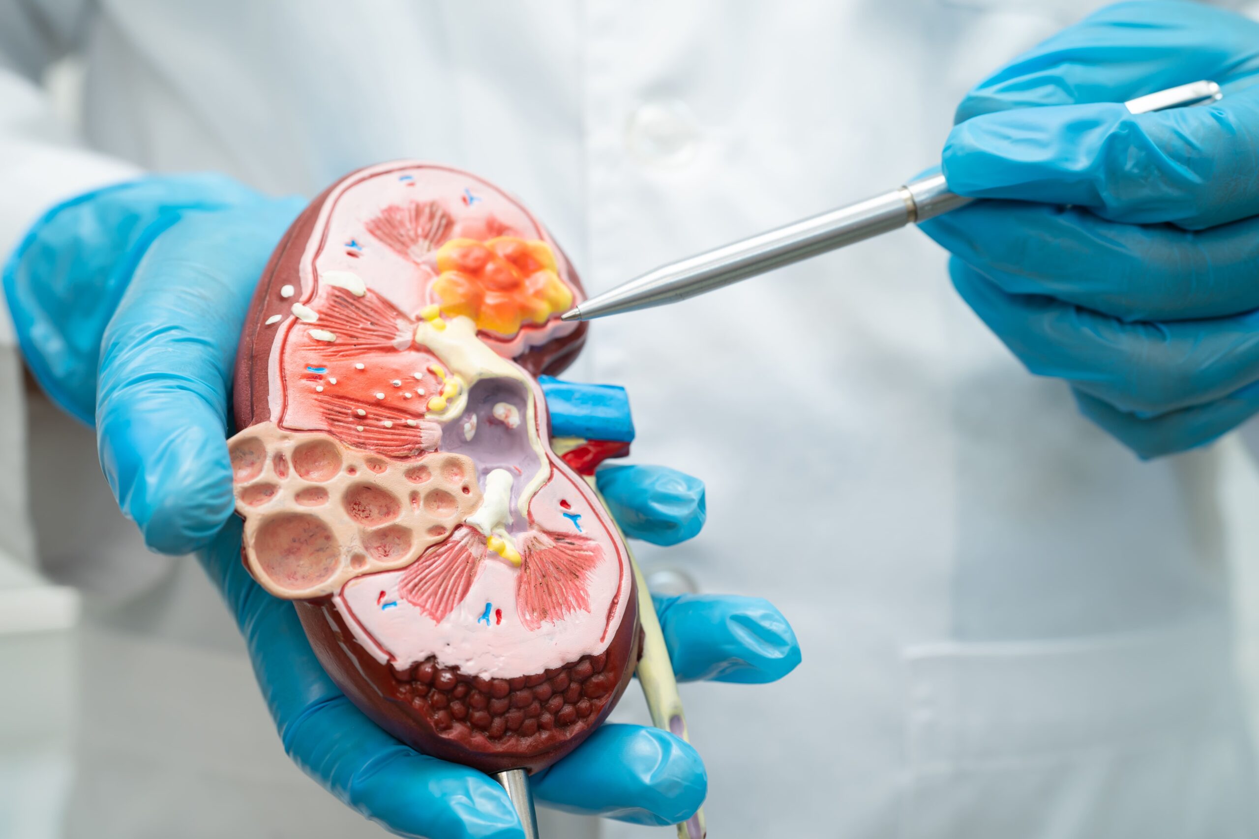
Bladder cancer staging (a system that describes how far cancer has spread) determines treatment approach and prognosis. Non-muscle-invasive bladder cancer (NMIBC) includes tumours confined to the inner lining (Ta). It also includes those invading the lamina propria (the layer of tissue beneath the bladder lining) (T1) and flat carcinoma in situ (CIS). These stages account for many newly diagnosed cases. They typically receive treatment via cystoscopy.
Muscle-invasive bladder cancer (MIBC) penetrates the detrusor muscle layer (the thick muscle layer that forms the bladder wall), comprising the bladder wall (T2). It extends through the muscle into the surrounding fat (T3) or invades adjacent organs, such as the prostate or uterus (T4). This distinction affects treatment planning. MIBC generally requires bladder removal or combined chemotherapy and radiation. Your healthcare provider can discuss a treatment plan tailored to your specific tumour characteristics, overall health status, and personal preferences.
Tumour grade describes cellular appearance under microscopy. Low-grade tumours contain cells that resemble normal urothelium and exhibit orderly growth patterns. High-grade tumours show disordered architecture with abnormal nuclei. This indicates aggressive behaviour regardless of stage. A small high-grade T1 tumour may pose a greater risk than a larger low-grade Ta tumour.
Transurethral resection of bladder tumour (TURBT) (a procedure where the surgeon removes tumours from the bladder using instruments passed through the urethra) serves dual diagnostic and therapeutic purposes. This procedure removes visible tumours through the cystoscope. It provides tissue for pathological staging while eliminating disease. Complete resection of non-muscle-invasive tumours may be curative. However, the risk of recurrence necessitates ongoing surveillance.
Intravesical therapy (treatment where medication is delivered directly into the bladder through a catheter) delivers medication directly into the bladder following TURBT. BCG immunotherapy (a treatment that uses weakened tuberculosis bacteria to stimulate the immune system) stimulates an immune attack against residual cancer cells. It helps reduce the risk of recurrence in high-risk NMIBC. Chemotherapy agents (cancer-killing drugs) such as mitomycin C may be instilled immediately after resection or as part of treatment courses for specific tumour profiles.
Surveillance cystoscopy follows initial treatment at intervals determined by tumour risk category. High-risk tumours require cystoscopy every three months initially. Intervals extend if no recurrence appears. Low-risk tumours permit less intensive surveillance but still require regular monitoring given recurrence rates. Upper tract imaging (scans of the kidneys and ureters) is periodically performed to screen for new ureteral or kidney tumours.
| Symptom | Characteristics | Urgency |
|---|---|---|
| Gross haematuria | Visible blood, any duration | Prompt evaluation |
| Microscopic haematuria | Found on urinalysis | Schedule consultation |
| Irritative symptoms without infection | Urgency, frequency, dysuria | Evaluation recommended |
| Combination symptoms | Multiple urinary changes | Priority assessment |
| Pain with haematuria | Flank or pelvic discomfort | Urgent evaluation |
Smoking cessation can provide significant risk reduction for current smokers. While accumulated damage doesn’t immediately reverse, quitting halts ongoing carcinogen exposure. It begins with a gradual decrease in risk. Support programmes, nicotine replacement, and prescription medications improve cessation success rates.
Adequate hydration dilutes urinary carcinogens and increases voiding frequency. This reduces contact time between harmful substances and the bladder lining. While specific fluid intake targets lack strong evidence, maintaining light-coloured urine suggests adequate hydration.
Occupational safety measures protect workers in high-risk industries. Proper protective equipment, ventilation systems, and exposure monitoring reduce exposure to carcinogens. Workers should maintain awareness of occupational health guidelines and report concerning exposures.
✅ Quick Tip
Keep a brief symptom diary if you notice urinary changes. Recording bleeding episodes, colour, associated symptoms, and timing helps your urologist identify patterns and determine appropriate investigation urgency.
Can bladder cancer symptoms come and go?
Yes, particularly haematuria. Bleeding from bladder tumours typically occurs intermittently rather than continuously. Blood may appear for several days, stop completely for weeks, then recur. Any episode of blood in urine warrants investigation, regardless of whether it continues.
Do bladder cancer symptoms differ between men and women?
The symptoms themselves are similar. However, women may experience delayed diagnosis because haematuria can be attributed to menstruation or urinary tract infections, both of which are common in women. Additionally, irritative symptoms in women may be initially treated as recurrent UTIs without further investigation. Women with persistent urinary symptoms despite negative cultures should receive urological evaluation.
How quickly do bladder tumours grow?
Growth rates vary considerably based on tumour grade and biology. Low-grade tumours may grow slowly over months to years, while high-grade tumours can progress rapidly. Carcinoma in situ can remain flat for extended periods before becoming invasive. This variability makes prompt evaluation of symptoms significant.
Can bladder cancer be detected through routine blood tests?
Standard blood tests don’t detect bladder cancer. However, they may show anaemia (low red blood cell count) from chronic bleeding. No widely available blood marker reliably screens for bladder cancer. Urinalysis detecting microscopic blood often provides the first laboratory indication prompting further investigation. Urine-based tumour markers (substances released by cancer cells that can be detected in urine) exist but aren’t recommended for routine screening (testing healthy people to detect potential problems early) in average-risk individuals.
What’s the difference between bladder infection and bladder cancer symptoms?
Both can cause urgency, frequency, and discomfort during urination. Bladder infections typically cause cloudy, foul-smelling urine and respond to antibiotics within days. Bladder cancer symptoms that persist despite treatment may include painless bleeding and worsen gradually. Recurrent “infections” with negative cultures or symptoms unresponsive to appropriate antibiotics should prompt cystoscopy.
Important information: Individual experiences with bladder cancer symptoms, diagnosis, and treatment vary based on personal health factors, tumour characteristics, and other medical conditions. The information provided here is educational and should not replace consultation with qualified healthcare professionals who can assess your specific situation and provide tailored advice.
Blood in urine requires investigation regardless of amount or duration. Urological evaluation determines whether bleeding originates from bladder tumours, infection, stones, or other causes. Persistent urinary changes without infection also warrant cystoscopy to exclude bladder pathology.
If you’re experiencing blood in urine, persistent urgency or frequency, or unexplained bladder discomfort, consult Dr Azhari for comprehensive evaluation and appropriate diagnostic testing.

Former Director of Endourology (Urinary stone service) Singapore General Hospital 2016 to 2023
With more than 20 years experience as a certified Urologist, Dr Nor Azhari specializes in treating a wide range of kidney, bladder and prostate conditions as well as disorders of the male reproductive organs. He offers minimally invasive treatment options and provides same-day appointments for convenience.
For urgent or same day appointment requests, please call our hotline.

Learn about Peyronie's disease treatment options in Singapore, from oral medications to surgery. Und

Adult circumcision in Singapore—medical reasons, surgical techniques, recovery timeline, and post-

How diabetes causes erectile dysfunction and management strategies. Urology guidance for diabetic me
Understand what to expect during a cystoscopy procedure, from preparation to recovery. Learn about b

Learn why recurrent UTI in men requires thorough investigation, underlying causes, diagnostic approa

Understand haematuria causes, from UTIs to bladder conditions. Learn when blood in urine needs medic

Understand kidney cyst causes, diagnosis methods, and when treatment may be needed from a urological

Dietary and lifestyle strategies to prevent kidney stones from recurring. Practical hydration, diet,
PCNL surgery removes large kidney stones through a small incision. Learn about the procedure, recove
Learn how ESWL kidney stone treatment works, who may be suitable, and what to expect. A urologist ex
 Optimized by Seraphinite Accelerator