Curvature of the Penis: Understanding Peyronie’s Disease and Treatment Options
Learn about Peyronie's disease treatment options in Singapore, from oral medications to surgery. Und

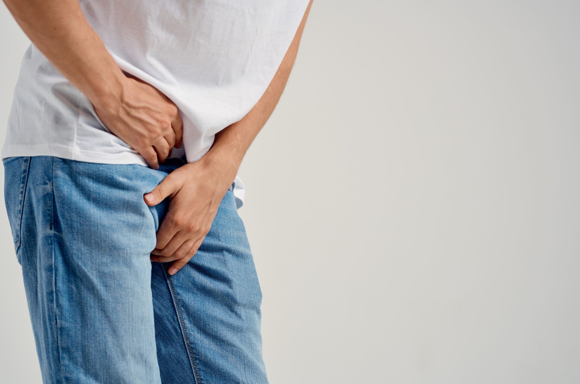
Can you identify whether a lump in your scrotum is benign or requires urgent medical attention? The scrotum contains multiple structures beyond the testicles themselves. These include the epididymis (a coiled tube that stores and transports sperm), the spermatic cord (which contains blood vessels, nerves, and the tube that carries sperm), and the surrounding tissue layers. Each structure can develop masses with distinct characteristics. Testicular lump pain, when present, provides diagnostic information.
However, painless masses also warrant evaluation in specific scenarios. Physical examination combined with ultrasound imaging (a non-invasive scan using sound waves to create images of internal structures) allows urologists—doctors who specialise in conditions affecting the urinary system and male reproductive organs—to characterise scrotal abnormalities and determine appropriate management.
The scrotum houses several distinct anatomical structures. Understanding their arrangement helps localise where lumps originate.
The testicle itself sits within the tunica vaginalis, a fluid-filled sac that allows the testicle to move freely. Behind and above each testicle lies the epididymis—a coiled tube storing and transporting sperm. The spermatic cord runs from the testicle upward through the inguinal canal. It contains blood vessels, nerves, and the vas deferens (the tube that carries sperm from the epididymis).
Lumps arising directly from testicular tissue carry different implications than those originating from surrounding structures. Epididymal cysts develop in the epididymis. You can distinguish them by their position relative to the testicle. Spermatic cord masses typically present higher in the scrotum, near the inguinal region.
During self-examination, these anatomical landmarks guide assessment. The testicle should feel smooth and oval-shaped, while the epididymis feels like a soft ridge along the back. Any complex, irregular, or fixed mass within the testicle itself requires prompt evaluation.
Epididymal cysts represent commonly encountered scrotal masses. These fluid-filled sacs develop within the epididymis. They typically feel smooth, round, and separate from the testicle. They transilluminate when a light shines through them. This is a diagnostic feature that distinguishes fluid-filled from solid masses.
Spermatoceles are similar cysts containing sperm-filled fluid. They are usually located at the head of the epididymis. Both conditions rarely cause symptoms unless they grow large enough to create a sensation of heaviness or mild discomfort.
Treatment becomes necessary only when cysts cause significant discomfort or grow to bothersome sizes. Surgical excision (a procedure where the doctor removes the cyst) remains the definitive option. However, aspiration (in which the doctor drains fluid with a needle) can provide temporary relief in select cases.
Hydroceles occur when fluid accumulates within the tunica vaginalis surrounding the testicle. The scrotum appears swollen, sometimes considerably so. However, the underlying testicle remains normal. Hydroceles typically develop gradually and cause minimal pain in the testicular lump. The weight and size may create discomfort.
Congenital hydroceles (those present from birth) in infants often resolve spontaneously as the communication with the abdominal cavity closes. Adult-onset hydroceles may develop following infection, injury, or without an identifiable cause. Large or symptomatic hydroceles may benefit from surgical repair via hydrocelectomy (a procedure in which the surgeon drains the fluid and may remove or alter the sac to prevent recurrence).
Varicoceles—dilated veins within the spermatic cord—are a known condition, particularly on the left side due to venous drainage patterns. They characteristically feel like a “bag of worms.” They become more prominent when standing or performing the Valsalva manoeuvre (bearing down as if having a bowel movement).
Varicoceles can represent a treatable cause of male infertility. The elevated scrotal temperature can impair sperm production.
Testicular torsion constitutes a urological emergency. The spermatic cord twists, cutting off the blood supply to the testicle. Symptoms appear suddenly. These include severe testicular lump pain, scrotal swelling, nausea, and sometimes abdominal discomfort. The affected testicle often sits higher than usual and may be oriented horizontally.
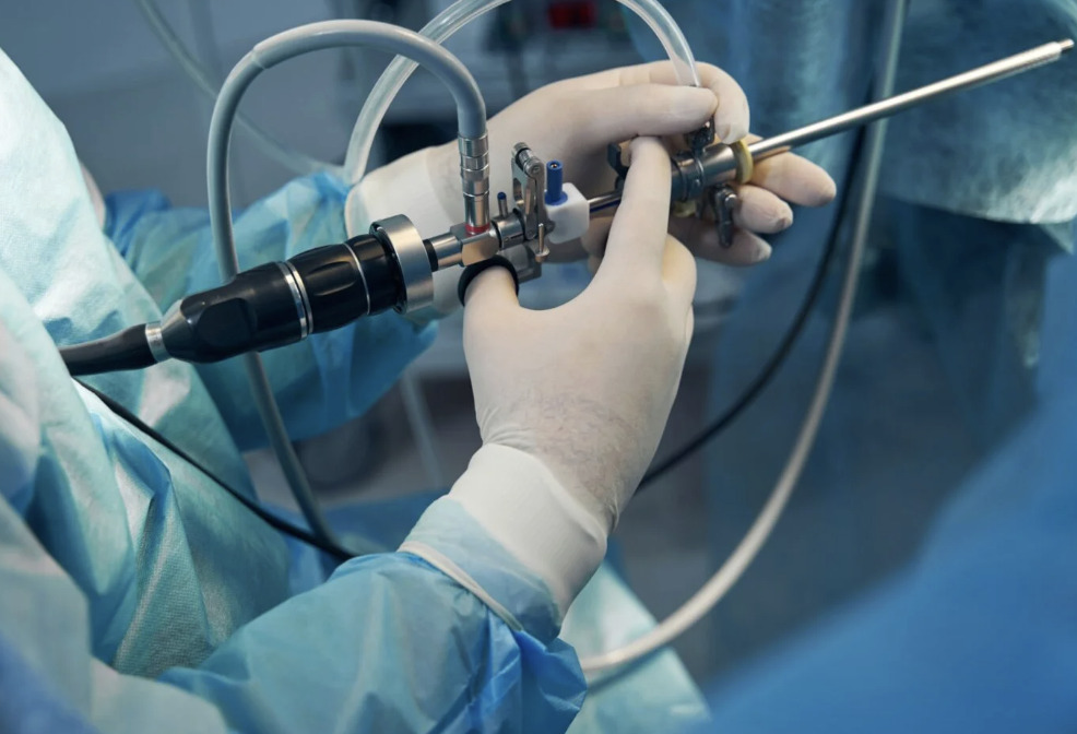
Time sensitivity is essential. Testicular salvage rates drop after several hours of torsion. Surgical exploration (where the surgeon opens the scrotum to examine the testicle) and detorsion (untwisting the spermatic cord) should occur promptly. The surgeon performs fixation of both testicles (stitching them in place) to prevent recurrence.
Young men and adolescents face a higher risk. However, torsion can occur at any age. Any sudden-onset severe testicular pain warrants immediate medical evaluation—not observation at home.
Epididymitis—infection or inflammation of the epididymis—causes gradual-onset scrotal pain that worsens over days. The epididymis feels swollen and tender. Fever may accompany the condition. In younger men, sexually transmitted infections (infections passed through sexual contact, such as chlamydia or gonorrhoea) commonly cause epididymitis. Older men are more likely to develop infections caused by urinary tract bacteria.
Orchitis refers to testicular inflammation. It frequently occurs alongside epididymitis (epididymo-orchitis). Viral orchitis, classically associated with mumps, can directly affect the testicle.
When initiated appropriately, antibiotic treatment can resolve bacterial infections. Supportive measures—scrotal elevation, ice application, and anti-inflammatory medications—provide symptomatic relief during recovery.
Testicular cancer typically presents as a painless, hard lump within the testicle itself. The lump is distinct from the surrounding structures. The mass feels fixed to the testicular tissue and may alter the testicle’s normal smooth contour. While testicular lump pain occurs in some cases, the absence of pain should not provide false reassurance. Timely treatment can improve outcomes.
Risk factors include:
Men in young adulthood face a higher incidence. However, the condition can develop at any age.
Ultrasound imaging (a scan using sound waves to create detailed images) characterises suspicious masses. Tumour markers—blood tests measuring specific proteins (AFP, beta-HCG, LDH) that may be elevated in testicular cancer—provide additional diagnostic information. Treatment typically involves orchiectomy (surgical removal of the affected testicle). Doctors follow this with surveillance or additional therapy based on staging (determining how far the cancer has spread).
? Did You Know?
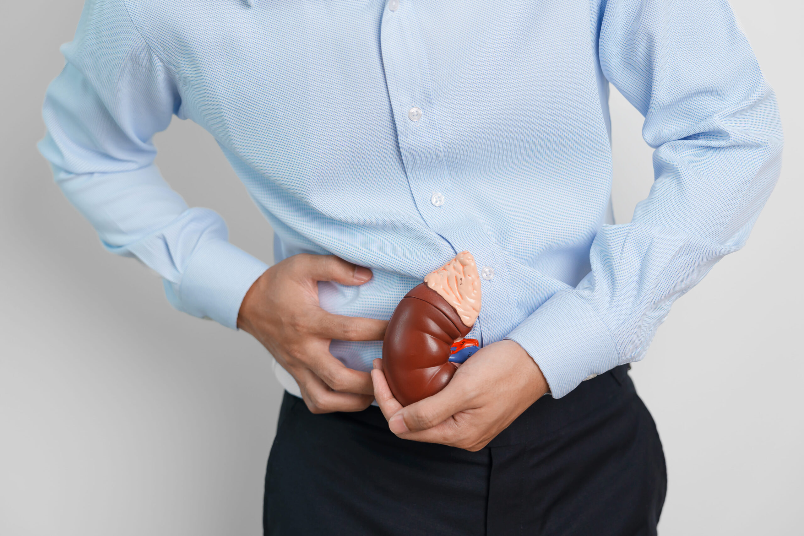
The testicles develop near the kidneys during foetal life and descend into the scrotum before birth. This migration explains why testicular pain sometimes radiates to the lower abdomen and why undescended testicles carry increased cancer risk.
Urological examination systematically evaluates scrotal contents to localise and characterise masses. Assessments include:
Examination while standing and lying down reveals varicoceles. They decompress in the supine position.
Scrotal ultrasound provides detailed visualisation of testicular and extratesticular structures. This non-invasive, radiation-free imaging modality (a type of scan) distinguishes:
Ultrasound findings guide management decisions. They determine whether additional investigations—such as tumour markers and CT staging (scans to check whether cancer has spread)—may be indicated.
Regular testicular self-examination supports early detection of changes. Examine during or after a warm shower. This relaxes the scrotal skin and facilitates assessment.
Examination steps:
Any new lump, change in consistency, or persistent discomfort merits professional evaluation. Monthly self-examination establishes familiarity with normal anatomy. This makes abnormalities more apparent.
⚠️ Important Note
Self-examination supplements rather than replaces professional evaluation. Any concerning finding should prompt consultation with a urologist rather than continued self-monitoring.
Can I tell whether a scrotal lump requires evaluation just by how it feels?
Certain features raise concern. These include hard consistency, irregular borders, fixation to the testicle, and painless presentation. However, physical characteristics alone don’t always definitively distinguish benign from malignant conditions. Ultrasound imaging provides characterisation. Any new scrotal mass should be assessed by a healthcare professional regardless of how it feels.
Should I be concerned if a scrotal lump doesn’t hurt?
Painless lumps warrant evaluation. While many benign conditions like epididymal cysts cause no discomfort, testicular cancer typically presents as a painless mass. The absence of testicular lump pain should not delay seeking medical assessment. Early evaluation ensures diagnosis regardless of symptom severity.
How quickly should I see a doctor for a new scrotal lump?
Sudden severe pain requires emergency evaluation within hours due to torsion risk. Qualified healthcare professionals should assess new painless lumps within one to two weeks. Gradual-onset pain with fever suggests infection requiring evaluation within days. When uncertain, earlier assessment is preferable.
Can epididymal cysts become cancerous?
Epididymal cysts remain benign and do not transform into cancer. These fluid-filled structures are separate from the testicular tissue where cancers develop. However, any new or changing mass requires evaluation to confirm its benign nature. Do not assume it represents a known cyst.
What happens during a scrotal ultrasound?
Qualified healthcare professionals apply gel to the scrotum and move a handheld probe across the skin surface during an ultrasound. The procedure should not be painful and requires no special preparation. Images display immediately. This allows same-day discussion of findings in most cases.
Any new scrotal lump requires professional evaluation to distinguish benign cysts from more serious conditions. Painless masses warrant the same attention as painful ones, as testicular cancer often presents without discomfort. Physical examination and ultrasound imaging provide definitive characterisation.
If you are experiencing a new scrotal lump, testicular pain, or changes in testicular consistency, consult Dr Azhari for a comprehensive evaluation and appropriate management.

Former Director of Endourology (Urinary stone service) Singapore General Hospital 2016 to 2023
With more than 20 years experience as a certified Urologist, Dr Nor Azhari specializes in treating a wide range of kidney, bladder and prostate conditions as well as disorders of the male reproductive organs. He offers minimally invasive treatment options and provides same-day appointments for convenience.
For urgent or same day appointment requests, please call our hotline.

Learn about Peyronie's disease treatment options in Singapore, from oral medications to surgery. Und

Adult circumcision in Singapore—medical reasons, surgical techniques, recovery timeline, and post-

How diabetes causes erectile dysfunction and management strategies. Urology guidance for diabetic me
Understand what to expect during a cystoscopy procedure, from preparation to recovery. Learn about b

Learn about bladder cancer symptoms and when to seek medical evaluation. A urologist explains warnin
Learn why recurrent UTI in men requires thorough investigation, underlying causes, diagnostic approa

Understand haematuria causes, from UTIs to bladder conditions. Learn when blood in urine needs medic
Understand kidney cyst causes, diagnosis methods, and when treatment may be needed from a urological

Dietary and lifestyle strategies to prevent kidney stones from recurring. Practical hydration, diet,
PCNL surgery removes large kidney stones through a small incision. Learn about the procedure, recove
 Optimized by Seraphinite Accelerator